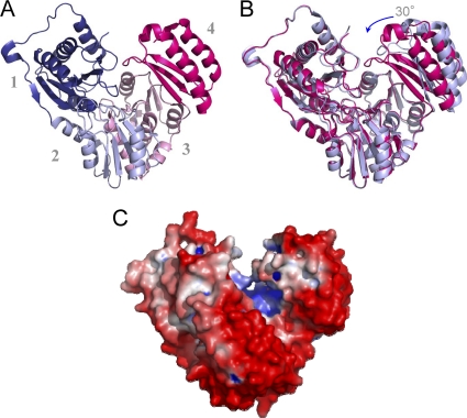
Fig. 2.
(A) A ribbon diagram of the BaPNGM monomer (chain B), showing its four domains in the following colors: domain 1 (dark blue; residues 1 to 152), domain 2 (light blue; residues 153 to 256), domain 3 (pink; residues 257 to 369), and domain 4 (magenta; residues 370 to 446). (B) A superposition of the two monomers in the asymmetric unit of the crystals. Monomer B (closed) is shown as magenta, and monomer A (open) is light blue. The rotation of domain 4 is indicated by an arrow. (C) The electrostatic potential of BaPNGM shown on a surface rendering. Negative charge is red, and positive charge is blue. The figure was made by using PMV, MSMS, and Chimera (22, 27, 28).

