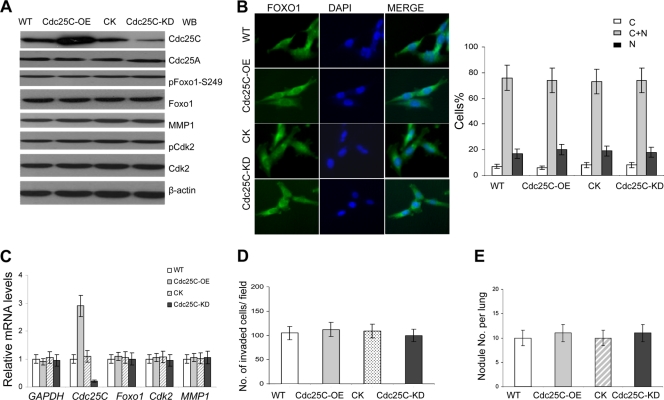
Fig. 10.
Altered Cdc25C expression does not affect the Fox1-MMP1 pathway and the metastatic phenotype of breast cancer cells. (A) Cdc25C was effectively and specifically overexpressed (Cdc25C-OE) or knocked down (Cdc25C-KD) in MDA-MB-231 breast cancer cells. The control Cdc25A levels were intact in the breast cancer cell lines. Lysates of the cancer cells were immunoblotted with the indicated antibodies. Total Foxo1 and MMP1 showed no significant changes in the cells with modulation of Cdc25C. (B) Increased or decreased Cdc25C expression (Cdc25C-OE or Cdc25C-KD) did not affect the cellular location and stability of Foxo1 in breast cancer cells. (Left) Foxo1. (Middle) DAPI. (Right) Merge. Quantification of Foxo1 localization in cells is shown in the bar graphs. n=200 for each cell type. (C) Foxo1 and MMP1 mRNA levels were evaluated in breast cancer cells with modulation of Cdc25C by quantitative real-time PCR; human GAPDH and Cdk2 served as loading controls (means ± SD; n=5). (D) Cell invasion assay on Matrigel-coated Transwells and statistical analysis of cell invasive ability in vitro. The cells that invaded the lower surface of the filter were counted in 10 random fields under a light microscope at high magnification (×100 [B]). The error bars represent the means ± SD; n=10. (E) Metastasis assay in vivo. The nodules per mouse lung for all 4 sections were counted under a light microscope (injected via the tail vein; 10 mice for each cell type). Similar results were also observed for the assay of MDA-MB-231 and MDA-MB-453 breast cancer cells versus their derivatives. The experimental evidence showed that Cdc25C did not regulate Foxo1-MMP1 and metastasis of these cancer cells, although altered Cdc25C expression affected the cell cycle through the G2/M checkpoint (11, 18).

