Fig. 4.
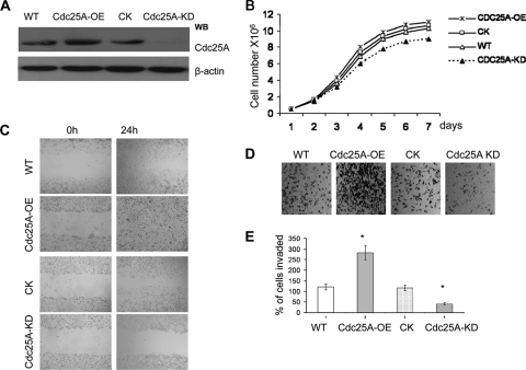
Cdc25A expression mediates breast cancer cell invasion phenotypes in vitro. (A) Cdc25A was effectively and specially overexpressed (Cdc25A-OE) or knocked down (Cdc25A-KD) in breast cancer cells. (B) Growth curve of MDA-MB-231 breast cancer cells and their derivatives Cdc25A-OE and Cdc25A-KD. The WT or cells with empty vector served as controls (mean ± SD; n=5). P > 0.5 for Cdc25A-OE compared with the control or WT cells; P < 0.05 for Cdc25A compared with the control or WT cells. (C) Wound-healing assays. (D) Cell invasion assay on Matrigel-coated Transwells. (E) Statistical analysis of cell invasive ability. The cells that invaded the lower surface of the filter were counted in 10 random fields under a light microscope at high magnification (×100 [D]). P < 0.01 compared with the control and WT cells. The images are representative of those for the assay of breast cancer MDA-MB-231 and MDA-MB-453 cells versus their derivatives.

