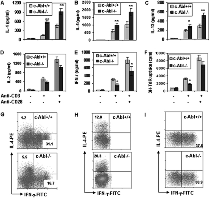
Fig. 1.
Cytokine production and proliferation of c-Abl−/− and wild-type T cells. (A to E) Naïve CD4+ CD25− CD44lo CD62Lhi T cells from c-Abl−/− and wild-type mice were isolated and stimulated with or without anti-CD3 (1 μg/ml) or anti-CD3 plus anti-CD28 (1 μg/ml) for 4 days. The production of IL-4 (A), IL-5 (B), IL-13 (C), IL-2 (D), and IFN-γ (E) was determined by ELISA. (F) The proliferation of c-Abl−/− and wild-type naïve T cells upon TCR/CD28 stimulation was examined by [3H]thymidine incorporation assay. Error bars represent data from three independent experiments. Student's t test was used for statistical analysis. *, P < 0.05; **, P < 0.01. (G) Naïve T cells from c-Abl+/+ and c-Abl−/− mice were stimulated with soluble anti-CD3 plus anti-CD28 for 5 days followed by an additional 4-hour stimulation with precoated anti-CD3 in the presence of brefeldin A (10 μg/ml). Intracellular staining for IL-4 and IFN-γ was performed, and cells were analyzed by fluorescence-activated cell sorting. (H and I) Naïve CD4+ T cells were cultured for 5 days under Th2 (1 μg/ml anti-CD3, 1 μg/ml anti-CD28, 5 μg/ml anti-IFN-γ, 5 μg/ml anti-IL-12, and 1 U/ml of IL-2) or Th1 (1 μg/ml anti-CD3, 1 μg/ml anti-CD28, 5 μg/ml anti-IL-4, and 1 U/ml of IL-2) polarization conditions, respectively. Cells were analyzed by intracellular staining and flow cytometry.

