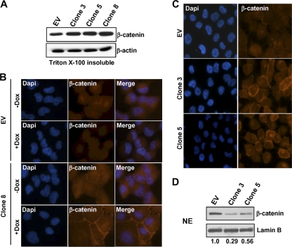
Fig. 7.
LOX-PP promotes β-catenin redistribution from the nucleus to the plasma membrane. (A) Control (EV) and LOX-PP-expressing clones 3, 5, and 8 were incubated in the presence of Dox for 48 h. The Triton X-100-insoluble membrane fraction was prepared and subjected to immunoblotting for β-catenin and for β-actin as a loading control. (B) Control (EV) or clone 8 H1299 lung cancer cells were incubated in the absence (−Dox) or presence (+Dox) of Dox for 48 h. For β-catenin staining, a primary mouse β-catenin antibody and a secondary anti-mouse Alexa Fluor 594 IgG antibody (red) were used. Nuclei were labeled with DAPI (blue). Fluorescence microscopy was performed on a Zeiss Axiovert 200 M microscope, and images taken using a 40× objective. Individual and merged images are shown. (C) Control (EV) or LOX-PP-expressing clones 3 and 5 were incubated in the presence of Dox for 48 h and subjected to fluorescence microscopy as described for panel B. (D) Control (EV) or LOX-PP-expressing clones 3 and 5 were incubated in the presence of Dox for 48 h, and nuclear extracts (NE) were prepared and subjected to immunoblotting for β-catenin and for lamin B, which confirmed essentially equal loading. This experiment and blots from two replicate experiments were scanned, and the changes relative to the results for the control EV, set at 1.0, are presented below.

