Fig. 1.
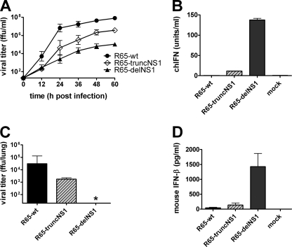
Growth and IFN induction of NS1 mutants of R65 in chicken embryo fibroblasts and mouse lungs. (A) CEF were infected with the indicated viruses at an MOI of 0.001. Virus titers in the cell culture supernatants were determined at the indicated times postinfection. Mean values and standard deviations (SD) of three independent experiments are shown. The origin of the y axis was set to the detection limit of the titration (101.3 FFU/ml). (B) CEF were infected with the various viruses at an MOI of 0.5 and analyzed for IFN activity in the supernatants at 24 h postinfection using a bioassay. The mean values and SD of three independent experiments are shown. (C and D) Mx1+/+ mice were infected intranasally with 5 × 10⁴ FFU of the indicated viruses per animal. At 24 h postinfection, the animals (n = 5 to 7) were killed, and virus titers (C) and IFN-β levels (D) in the lungs were determined by titration on MDCK cells and ELISA, respectively. The asterisk indicates that the viral titer in the sample was below the detection limit (101.3 FFU/ml).

