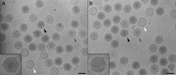
Fig. 4.
Cryo-electron microscopy of tagged capsids. Shown are micrographs of UL17-GFP (A) and wild-type (B) capsids. DNA-filled C capsids (black arrowheads and inset close-ups) are darker than, and easily distinguishable from, subset populations of A and B capsids (white and gray arrowheads, respectively). C capsids selected from each data set were subject to the three-dimensional reconstruction procedure.

