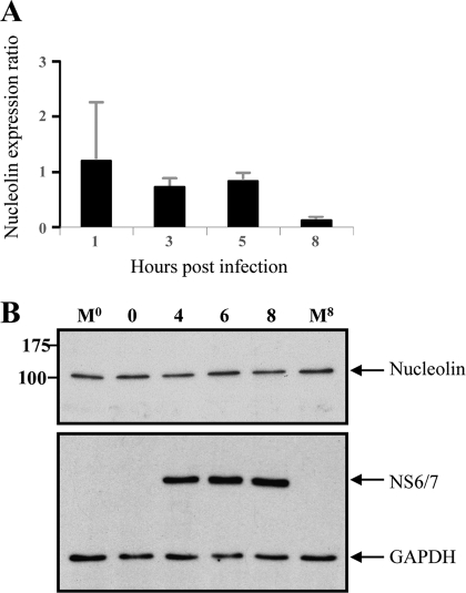
Fig. 3.
Nucleolin RNA and protein expression during FCV infection. (A) Total RNA extracted from FCV-infected CrFK cells at 1, 3, 5, and 8 h was subjected to quantitative RT-PCR using specific primers. Levels of expression of the nucleolin gene were obtained by relative quantification of the gene transcript, and expression of the GAPDH gene was used as a housekeeping gene control. Error bars show standard deviations. (B) Total cellular extracts from uninfected cells prepared at either time zero or 8 h (M0 and M8) and from FCV cells infected at an MOI of 3, obtained at 2, 4, 6, and 8 hpi, were analyzed by Western blotting using human antinucleolin (upper panel) or anti-FCV NS6/7 (lower panel) antibody. GAPDH was used as the loading control. Molecular sizes, in kDa, are shown on the left.

