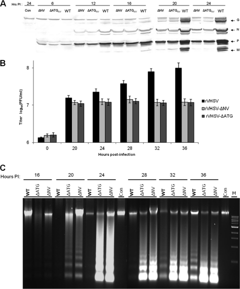
Fig. 5.
Replication and apoptotic kinetics of rVHSV, rVHSV-ΔNV, and rVHSV-ΔATG at the early stage of virus infection. EPC cells (∼5 × 10⁶) were inoculated with equal amounts of rVHSV, rVHSV-ΔNV, and rVHSV-ΔATG at an MOI of 1.0 PFU per cell. (A) Cell lysates were harvested and analyzed for the virus-specific proteins by immunoblotting. (B) Infected cells were freeze-thawed, and viruses were quantified by plaque assay at the indicated time points. (C) The low-molecular-weight DNAs from cell lysates were extracted and analyzed by a DNA laddering assay. Con, control.

