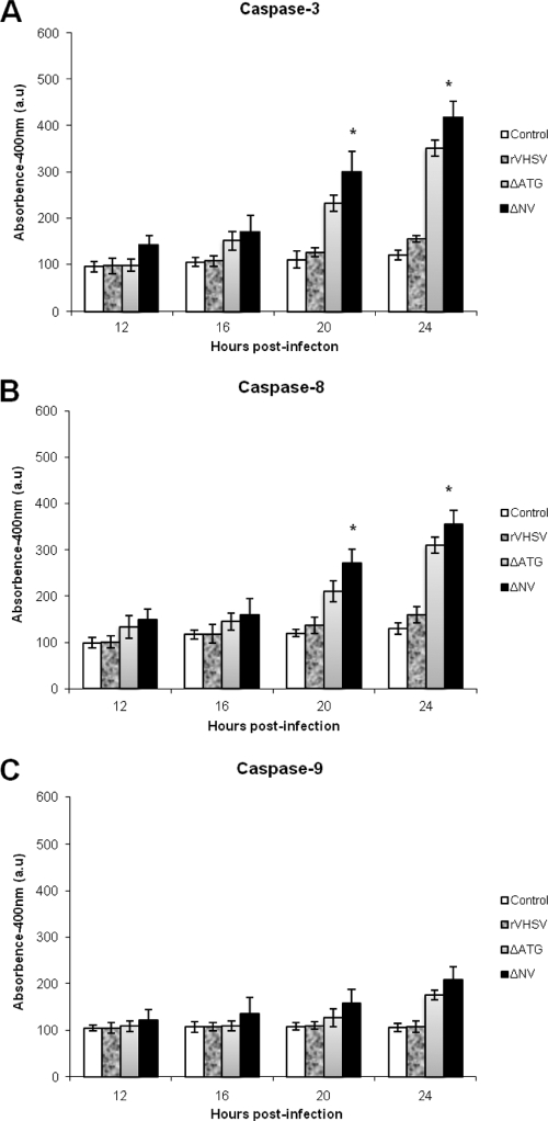
Fig. 6.
Caspase 3, 8, and 9 activities in rVHSV-, rVHSV-ΔNV-, and rVHSV-ΔATG-infected cells. EPC cells (∼3 × 10⁶) were inoculated with equal amounts of rVHSV, rVHSV-ΔNV, and rVHSV-ΔATG at an MOI of 1.0 PFU per cell, and cell lysates were prepared at the indicated times postinfection. Caspase 3-like (A), caspase 8-like (B), and caspase 9-like (C) activities at the indicated times after infection. All assays were performed as described in Materials and Methods (*, P < 0.05 compared to that of rVHSV-ΔATG).

