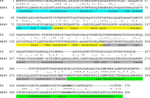
Fig. 2.
Alignment of bacteriophage T4 and RB49 Hoc sequences. β-Strands and α-helices are marked with the letters s and h, respectively. Regions corresponding to domains 1, 2, 3, and 4 of RB49 Hoc are shown in cyan, yellow, gray, and green, respectively. Since the structure of domain 4 of Hoc was not available, the secondary structure for this domain was predicted using the JPRED3 server (http://www.compbio.dundee.ac.uk/www-jpred) (7). The conserved motif in domain 4 is shown in boldface.

