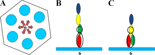
Fig. 6.
Schematic representation of Hoc bound to a gp23 hexamer. (A) The gp23 capsomer is represented by a hexagon. Six blue circles represent the gp23 monomers. The six possible orientations of the Hoc molecule with respect to the gp23 capsomer are represented by arrows. (B and C) The gp23 capsomer is represented by the blue bar. Domains 1, 2, 3, and 4 of Hoc are represented by blue, yellow, green, and red ellipses, respectively. The dumbbell-shaped density of Hoc, observed in the cryo-EM reconstruction, is represented by the black ellipse and circle. (B) Elongated conformation of the Hoc molecule. Domains 1 and 2 are presumably not visible in the cryo-EM density, in part because of a likely different displacement of the Hoc domains from the 6-fold axis of the gp23 hexamer in different particles used to generate the cryo-EM density. (C) Alternative structure of Hoc in which domains 2, 3, and 4 contribute to the observed cryo-EM density.

