Fig. 1.
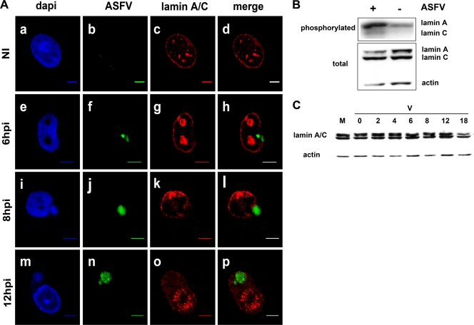
Peripheral lamin A/C phosphorylation and disruption at early times in ASFV infection. (A) 3D immuno-FISH of Vero cells uninfected (NI) and infected at 6, 8, and 12 h p.i. with 5 PFU of the Ba71V strain of ASFV. Nuclei were counterstained with DAPI (blue signal in a, e, i, and m), labeled with the FITC-conjugated ASFV genome probe (green signal in b, f, j, and n), and stained with a monoclonal antibody against lamin A/C (red signal in c, g, k, and o). Scale bars, 5 μm. (B) ASFV infection induces increased phosphorylation of lamin A and C in Vero cells. (Top) Autoradiography showing the immunoprecipitated 32P-labeled lamin A/C proteins obtained from mock-infected (−) and ASFV-infected (+) Vero cells at 4 h p.i. (Bottom) Western blot analysis showing the total amounts of lamin A/C and β-actin contained in each cell extract. (C) Degradation of nuclear lamin A/C at late times after ASFV infection. Western blot analysis of cell extracts from mock-infected (M) and ASFV-infected (V) Vero cells at different times postinfection (0, 2, 4, 6, 8, 12, and 18 h p.i.) using a polyclonal antibody against nuclear lamin A/C and an anti-β-actin antibody as an endogenous control.

