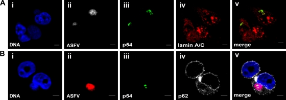
Fig. 2.
Localization of nuclear envelope markers in viral factories at 12 h p.i. with ASFV. Shown is 3D double immuno-FISH of Vero cells infected at 12 h p.i. with 5 PFU of the BA71V strain of ASFV. FISH was performed using the ASFV probe, followed by double IF using antibodies against nuclear envelope proteins and the ASFV protein p54 to label viral factories. (A) Lamin A/C localization in viral factories. (i) DAPI-counterstained DNA is shown in blue. (ii) ASFV DNA was visualized with streptavidin-TRITC, shown in gray. (iii and iv) 3D double immunofluorescence was developed using a polyclonal antibody against the ASFV marker p54, revealed with anti-rabbit-FITC (green signal in iii) and a monoclonal antibody against lamin A/C revealed with anti-mouse-Cy5 (red signal in iv). (v) Colocalization of both markers (yellow signal). (B) Nucleoporin p62 localization in viral factories. (i) DAPI-counterstained DNA is shown in blue. (ii) ASFV DNA was visualized with streptavidin-TRITC, shown in red. (iii and iv) 3D double immunofluorescence was developed using a polyclonal antibody against the ASFV marker p54, revealed with anti-rabbit-FITC (green signal in iii) and a monoclonal antibody against nucleoporin p62 revealed with anti-mouse-Cy5 (gray signal in iv). (v) Merged image. Scale bars, 5 μm.

