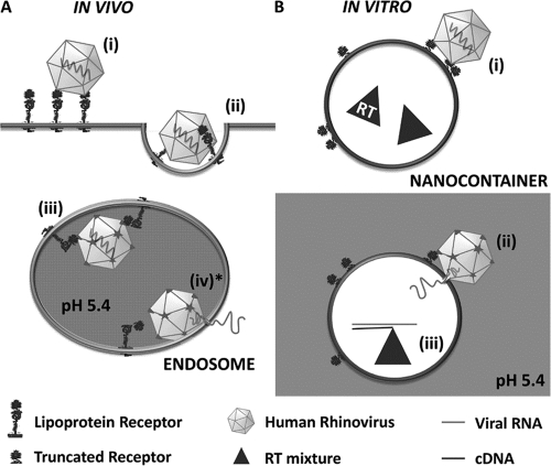
Fig. 1.
Scheme of in vivo and in vitro HRV2 uncoating. (A) In vivo uncoating in the cell. Virus binds to the receptor on the host cell (i) and is taken up via clathrin-dependent endocytosis (ii). Upon arrival in endosomes (iii), the low pH triggers virus release from its receptor. VP4 molecules and N-terminal sequences of VP1 are externalized, and low-pH-induced conformational changes of the receptor cause handing over of the hydrophobic particle to the lipid membrane. The hydrophobic domains of VP1 are inserted into the membrane and, most probably assisted by VP4, a pore is formed allowing for transfer of the RNA into the cytosol (iv*). (B) In vitro uncoating on nanocontainers. Virus is bound to truncated recombinant receptors attached via His6 tags to Ni2+-loaded NTA lipids on the surfaces of nanocontainers (i). Upon triggering the structural changes by exposure to acidic buffer, the RNA is transferred through the liposomal membrane into the aqueous lumen (ii), where it is transcribed into cDNA (iii) that is subsequently detected via PCR. Note that in vivo uncoating occurs from within whereas in the in vitro system it is from without. The step modeled experimentally in the present communication is marked with an asterisk.

