Fig. 4.
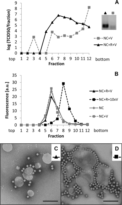
About 3 virions per liposome are sufficient to detect RNA transfer. Nanocontainers (NC) containing a fluorescent tracer lipid were decorated with receptor (R) and virus (V) was attached. Unbound particles were removed by flotation on a sucrose step gradient. (A) Infectious virus in the recovered fractions was detected via its infectivity as TCID50. (B) Nanocontainer-containing fractions were identified by fluorescence measurement. Binding of about 3 virus particles per liposome (i.e., resulting in a virus-to-lipid ratio of about 1.4 × 10⁻⁶) (see corresponding TEM image in panel C) led to slight broadening of the liposome peak. Higher virus concentrations (about 30 particles per liposome, i.e., at a virus-to-lipid ratio of about 1.4 × 10⁻⁵; see panel D) caused a shift of the liposome peak to higher density fractions. Coflotation of HRV2 occurred only with receptor-decorated vesicles; in the absence of the receptor, virtually all virus remained in the bottom fraction of the gradient (note the logarithmic scale) and no signal was seen for the RNA transfer reaction (A, inset). (C and D) Material in the pooled peak fractions in panel B was adsorbed to glow discharged, carbon-coated copper grids and stained with 2% phosphotungstate, pH 7.2. Images were taken under a transmission electron microscope at 5.4 × 10⁴-fold magnification. Size bar = 200 nm. Note that the faint band in the sample without receptor (inset) is irrelevant, as it was also present in samples without a template.

