
Fig. 3.
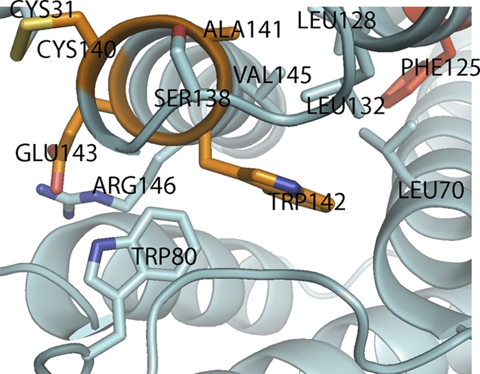
The CAWE motif of IFNφ2. Conserved residues in type I interferons in the vicinity of the CAWE motif are shown as sticks. The residues in the CAWE motif are colored orange. The highly conserved aromatic residue (Phe) is colored red. Visible helices and loops are labeled. See text for details.