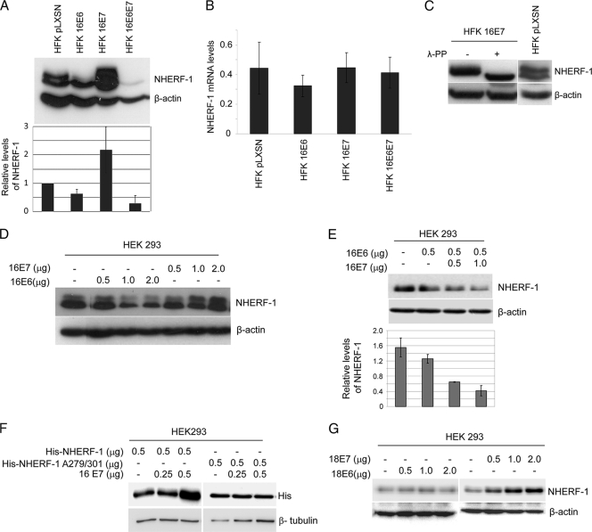
Fig. 3.
HPV16 E6 and E7 cooperate in NHERF-1 degradation. (A) Protein extracts of primary keratinocytes transduced with empty retrovirus vector (HFK pLXSN) or recombinant retroviruses, as indicated, were analyzed by immunoblotting using anti-NHERF-1 and anti-β-actin antibodies (top). The intensities of the protein bands in three independent experiments were quantified (bottom). (B) Total RNA was also extracted from the indicated cells, and NHERF-1 or GAPDH mRNA levels were measured by quantitative RT-PCR. y axis numbers represent arbitrary units of NHERF-1 mRNA levels in indicated cells standardized to the GAPDH levels. The data are the means of three independent experiments. The differences between the NHERF-1 levels in the different cells are not statically significant. (C) Protein extracts from HFKs transduced with empty (pLXSN) or HPV16 E7 retrovirus were incubated in the presence and absence of λ-PP and analyzed by immunoblotting using anti-NHERF-1 and anti-β-actin antibodies. (D) HEK293 cells were transfected with increasing concentrations of pLXSN HPV16 E6 or E7 as indicated. After 24 h, protein extracts were analyzed by immunoblotting. (E) HEK293 cells were transfected with pLXSN expressing HPV16 E6 together with increasing concentrations of pLXSN expressing HPV16 E7 as indicated. After 24 h, protein extracts were analyzed by immunoblotting (top). The intensities of the bands were quantified in three independent experiments and are represented in the histogram (bottom). (F) HEK293 cells were transfected with pcDNA3 expressing His-tagged NHERF-1 (His-NHERF-1), wild type or A279/A301 mutant, together with increasing concentrations of pLSXN expressing HPV16 E7 as indicated. After 24 h, protein extracts were analyzed by immunoblotting. (G) HEK293 cells were transfected with increasing concentrations of pLXSN expressing HPV18 E6 or E7 as indicated. After 24 h, protein extracts were analyzed by immunoblotting.

