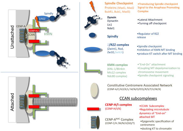
Figure 1.
Kinetochores have two structures depending upon whether they are attached to microtubules. The centromere/kinetochore can be subdivided into functional groups and biochemical subcomplexes, which are summarized according to the proteins in the subcomplexes and their major functions. Only the complexes discussed in this review are depicted. Unattached kinetochores (KT), which are assembled upon the constitutive centromere, recruit complexes with MT binding functions (Dynein and KMN) and the components of the mitotic checkpoint, which are thought to sense both occupancy and tensions across the centromere as a measure of proper microtubule attachment of the kinetochore. End-on microtubule attachment results in changes in the protein complexes present at the kinetochores. The constitutive centromere and components of the KMN network are retained; however, the RZZ complex, spindly, dynein and the mitotic checkpoint proteins are removed from the kinetochores upon end-on attachments (for a complete review see [5] [4]).

