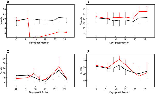
Figure 1.
Average percentage of peripheral lymphocyte subpopulations in cattle during experimental infection. Lymphocyte subpopulations of CD4+ (A), CD8+ (B), γδ+ (C), and B+ (D) are displayed. Values were generated using FACS staining of PBMCs from ten CD4+ T cell depleted (red) and ten control cattle (black) employing monoclonal antibodies specific for corresponding leucocyte markers. Standard deviations are displayed as bars.

