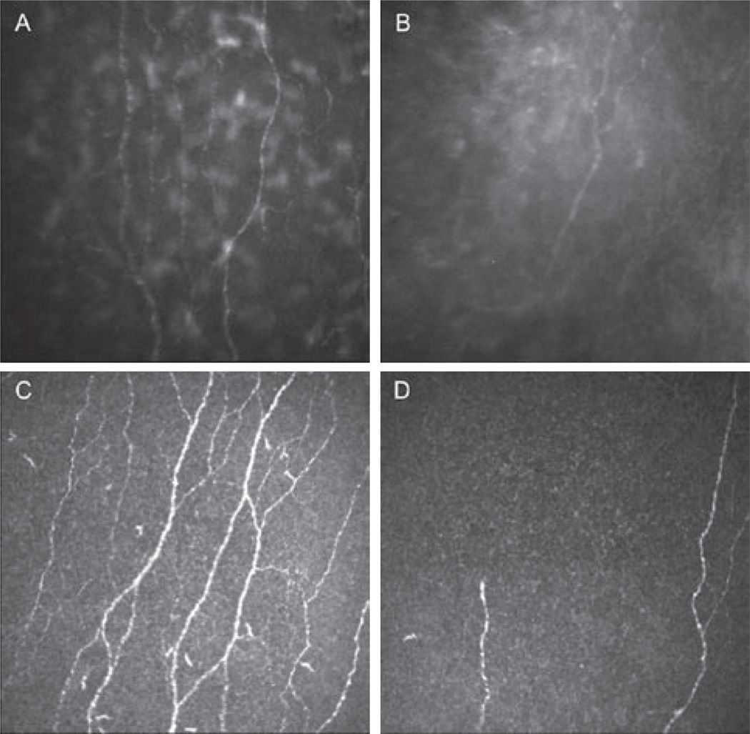
FIGURE 1.
Normal corneal sub-basal nerve plexus and after herpes simplex keratitis with two types of confocal microscopes. (A) Normal sub-basal nerve plexus. Slit scanning confocal microscopy (SSCM) Confoscan 4. (B) Herpes simplex keratitis with severe sensation loss. Note the decrease in total nerve count, length, and branching. SSCM. (C) Normal sub-basal nerve plexus. Laser scanning confocal microscopy (LSCM) HRT3/RCM. (D) Herpes simplex keratitis with severe sensation loss. Note the decrease in nerves density and branching. LSCM.

