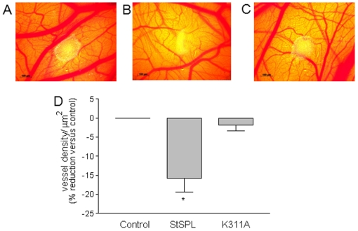
Figure 8. In vivo effect of StSPL on angiogenesis in the chicken chorioallantoic membrane (CAM).
MCF-7 cell spheroids containing 5×10⁵ cells in 50 µl were placed on E8 CAMs, and either treated with PBS (control), WT StSPL (StSPL, 20 µg/ml), or K311A (20 µg/ml) for 4 d. CAMs were analysed for vessel formation as described in the Materials and Methods section, and the density of vessels per µm2 of area around the tumor was determined. Representative CAMs that were PBS-treated (A), WT StSPL-treated (B), and K311A mutant-treated (C) were photographed under a stereomicroscope and vessel density was determined using the Vessel_tracer software [58] (D). Results are expressed as vessel density per µm2 and are means +/− S.D. (n = 5). *p<0.05 considered statistically significant when compared to the control treated samples.

