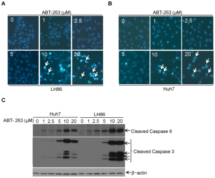
Figure 2. HCC cells are resistant to low doses of ABT-263.
A. LH86 and B. Huh7 cells were treated with ABT-263 (0–20 µM) for up to 24 h. Apoptosis was measured through Hoechst staining to show apoptotic cells with condensed nuclei as described in ‘materials and methods’. (representative apoptotic cells were marked with white arrows in ABT-263 treatment panel). C. HCC cells were treated with increasing doses of ABT-263 as indicated for up to 24 h. Then cells were harvested and cell lysates were prepared and subjected to Western blotting. Caspase activation was assessed through detecting the cleaved bands of caspase 9 and caspase 3. β-actin protein levels were used as an equal protein loading control.

