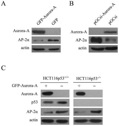
Figure 2. Regulation of AP-2α gene expression by Aurora-A.
(A) AP-2α protein levels in KYSE150/GFP-Aurora-A and KYSE150/GFP cells were analyzed using immunoblotting assay. The actin levels were measured as loading control. (B) AP-2α protein levels in EC9706/pGCsi???EC9706/pGCsi-Aurora-A cells were examined by Western Blotting assay. (C) The HCT116 p53+/+ and HCT116 p53-/- cells were transiently transfected with pEGFP-Aurora-A and control plasmids. Cells were lysed after 48h of incubation and evaluated for the expression of AP-2α by Western Blotting assay.

