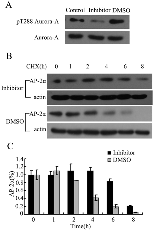
Figure 4. Aurora-A-mediated AP-2α degradation depends on Aurora-A kinase activity.
(A) KYSE150/GFP-Aurora-A cells were exposed to 1 µM of Aurora-A kinase inhibitor and cellular proteins were collected 2 hours later. Western Blot was performed with antibodies to pThr288 on Aurora-A or total Aurora-A. DMSO was used as a negative control. (B) KYSE150/GFP-Aurora-A cells were exposed to Aurora-A kinase inhibitor or DMSO for 2 hours prior to treatment with CHX. Cells were harvested at the indicated time points and analyzed by Western Blot. (C) The amounts of AP-2α were calculated by densitometry and normalized to corresponding actin levels. The column diagram represents the amount of normalized AP-2α at each time point comparing with the original levels (0 h).

