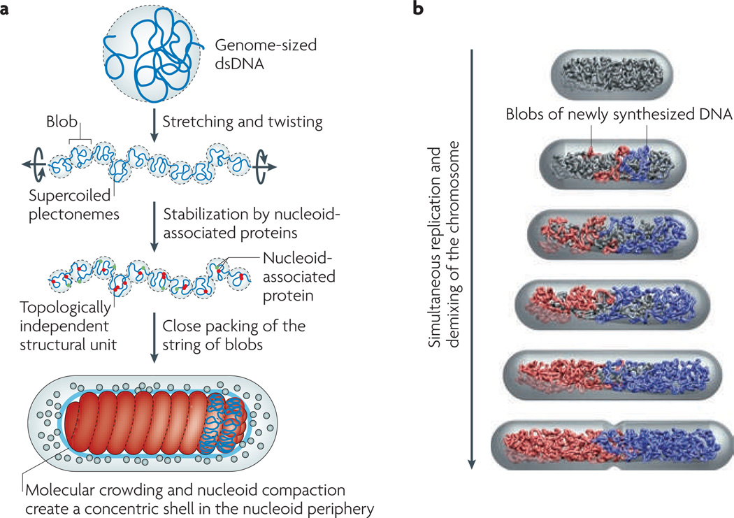
Figure 2. Physical model of a bacterial chromosome and its segregation.
a | A reductionist model of the Escherichia coli chromosome. First, we stretch a bacterial-genome-sized naked double-stranded DNA (dsDNA). This breaks the DNA into a series of blobs, the total volume of which gradually decreases as pulling continues. In parallel, we also twist the DNA to match the supercoil density of a bacterial chromosome. As a result, the DNA blobs will consist of supercoiled plectonemes (a shape of the DNA in which the two strands are intertwined). We stop the simultaneous pulling and twisting processes when the total volume of the blobs equals the target volume of the nucleoid inside the cell. Next, we ‘sprinkle’ the chromosome with nucleoid-associated proteins. These stabilize the supercoiled DNA blobs as topologically independent structural units of the chromosome. Finally, we connect the two ends of the chromosome to make it circular, and then pack it tightly in the cell. For the simpler case of chains without supercoiling, the phase diagram in FIG. 1 provides a model for the close-packed organization and segregatability of the chains inside the cell. In general, supercoiling will only increase the tendency for chromosome demixing because of the branched structure that it induces. b | The concentric shell model predicts extrusion of the newly synthesized DNA (blue and red) to the periphery of the nucleoid15. The newly replicated DNA is extruded to the periphery of the unreplicated nucleoid (grey) and forms a string of DNA blobs in the order of replication, promoted by SMC (structural maintenance of chromosomes) proteins and other nucleoid-associated proteins. In our model, the two strings of blobs repel each other and drift apart owing to the excluded-volume interaction and conformational entropy.

