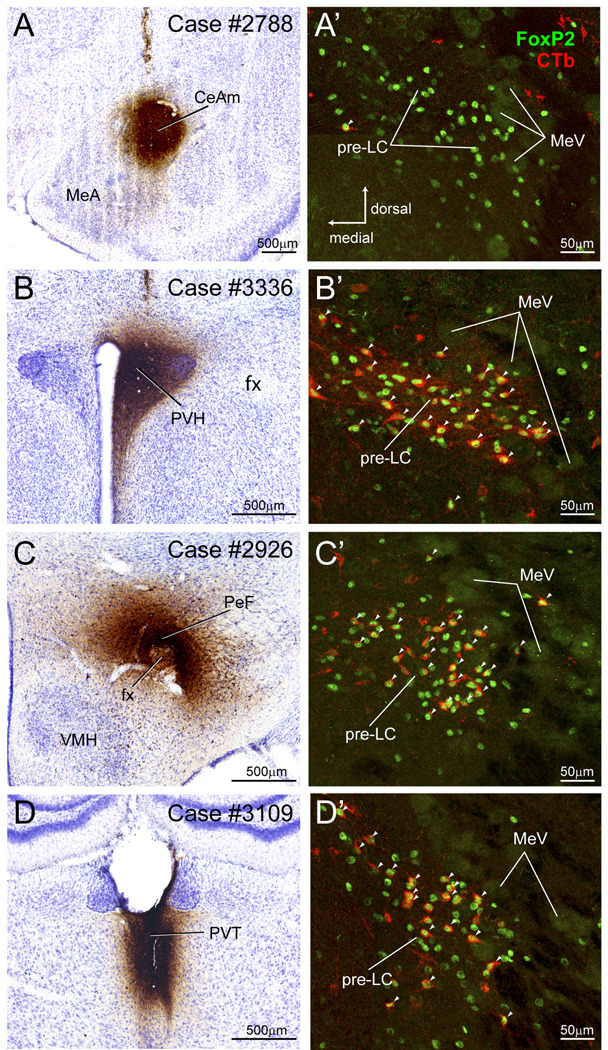
Figure 4.
Four CTb cases used to analyze whether FoxP2+ neurons in the pre-LC and PBel-inner project to the amygdala (A), hypothalamus (B and C), and thalamus (D). A. CTb injection in medial part of the central amygdaloid nucleus (CeAm). A’. FoxP2+ neurons in the pre-LC did not co-label with CTb indicating they do not project to the CeAm. As shown in Fig. 7, CTb labeled neurons were found in the PBel-inner, but none of them were FoxP2+. B. CTb injection in paraventricular hypothalamic nucleus (PVH). B’ Co-labeled CTb and FoxP2+ neurons in the pre-LC (Co-labeled neurons indicated by arrowheads). See Fig. 8B. C. CTb injection in the perifornical hypothalamic region. C’. Co-labeled CTb and FoxP2+ neurons in the pre-LC. See Fig. 9A. D. CTb injection in the paraventricular thalamic nucleus. D’. Co-labeled CTb and FoxP2+ neurons in the pre-LC. See Fig. 11A.

