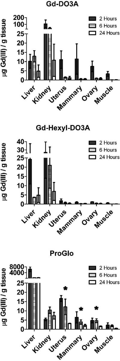
Figure 3.
Tissue distribution of Gd-DO3A (top), Gd-Hexyl-DO3A (middle), and ProGlo (bottom) in female CD-1 mice 2, 6, and 24 hours after injection. The levels of Gd(III) in the PR-rich tissues (uterus, ovaries, and mammary tissues) were significantly higher than in the muscle (which served as a negative control due to its low PR expression) at all time points after injection of ProGlo (Student's t test, p < 0.05). The levels of Gd(III) in the PR-rich tissues after injection of ProGlo were significantly higher than the levels after injection of Gd-DO3A after 2 hours (Student's t test, p < 0.05). Asterisks designate high retention of ProGlo in PR-rich tissues. Data are mean ± standard deviation.

