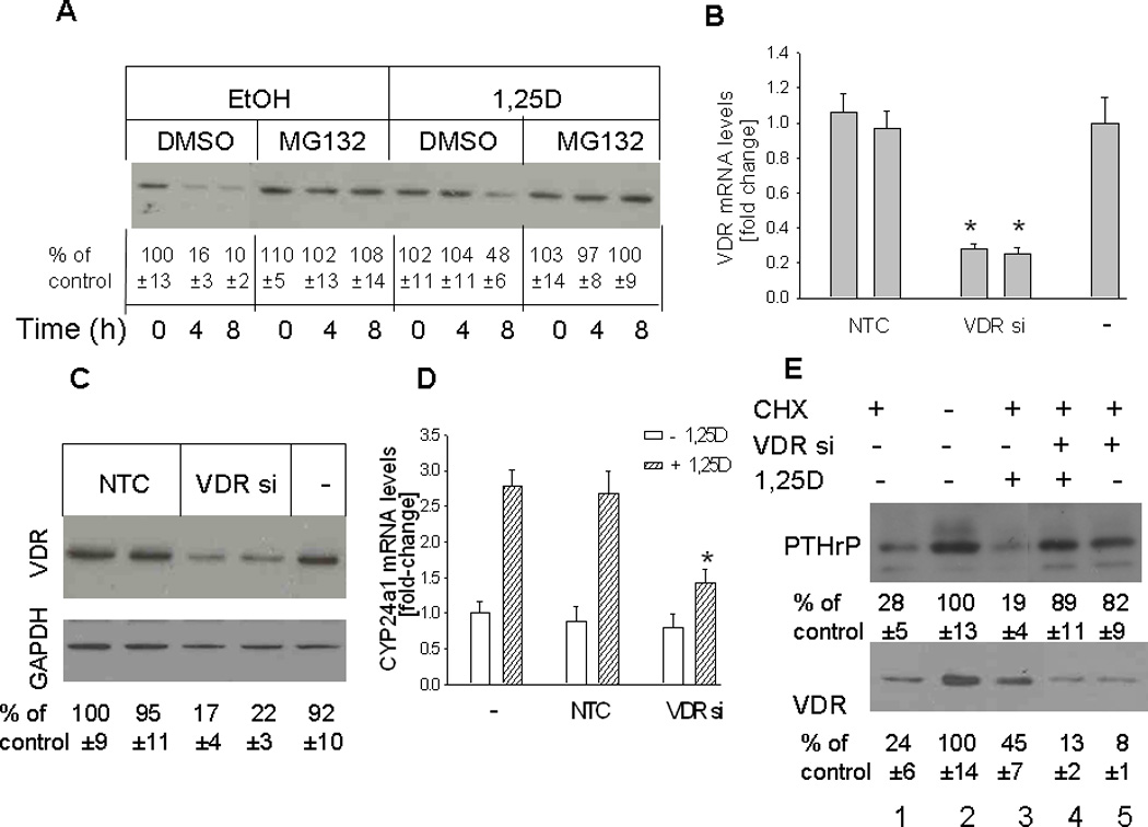
Figure 5.

(A) Time course for VDR depletion in cells treated with 1,25D or ethanol (vehicle control) Cells were pretreated with the protein synthesis inhibitor cycloheximide (CHX) for 30 min, then with 1,25D (10 −7M) in the presence or absence of the proteasome inhibitor MG132 (50 µM). Ethanol (EtOH) and DMSO were used as vehicle controls for 1,25D and MG132, respectively. Nuclear extracts were prepared at the indicated time points for Western blotting. (B–D) Effects of suppressing VDR expression on levels of the VDR (B,C) and of the 1,25D target gene CYP24a1 (D). VDR expression was suppressed by transfection with siRNA targeting the VDR. (B, D) VDR and CYP24a1 mRNA levels were analyzed by reverse transcription/real-time PCR analysis. Each bar is the mean ± SEM of three experiments for each of two independent non-target control siRNAs (NTC), VDR-targeting siRNAs (VDR si), or non-transfected control (−). (C) Western blot analysis for VDR levels in VDR siRNA-transfected cells. (E) Recovery of PTHrP expression in cells with suppressed VDR expression treated with 1,25D or ethanol. PC-3 cells transfected with an siRNA targeting the VDR (VDR si +) were pre-treated with the protein synthesis inhibitor cycloheximide (CHX) for 6 h, then with 1,25D (10−7 M) (+ 1,25D lanes) or with ethanol (vehicle control; − 1,25D lanes). − VDR si lanes were transfected with NTC siRNA. After 48 h, lysates were prepared for Western blotting. In (A), (C) and (E), the relative PTHrP levels were obtained after densitometric scanning of the Western blots and normalization to GAPDH. The control value (−, ethanol treated cells) was set at 100%. In (A), (C) and (E), the mean and S.E.M. values represent data from three independent experiments.
