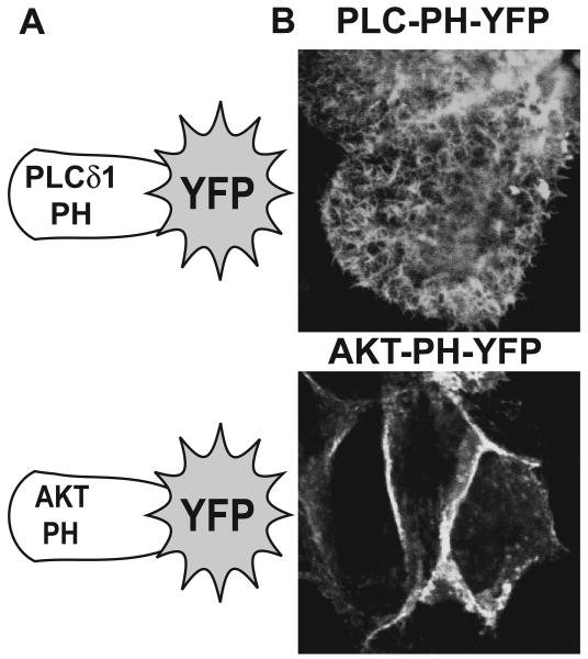
Figure 1.
PIP2 and PIP3 predominantly localize to apical and basolateral membranes of LLC-PK1 cells, respectively. A. Schematics depicting the pleckstrin homology domains from PLCδ1 and AKT fused to YFP for the intracellular detection of PIP2 and PIP3, respectively. B. LLC-PK1 cells plated on culture slides were transduced with adenoviruses expressing either PLC-PH-YFP (PIP2 sensor, upper panel) or AKT-PH-YFP (PIP3 sensor, lower panel). Representative confocal images of the apical domains (PLC-PH-YFP, upper panel) or basolateral domains (AKT-PH, lower panel) are shown.

