Figure 5.
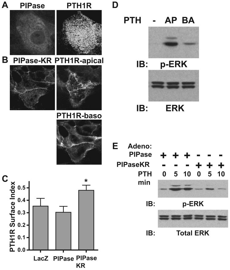
PIP2 depletion disrupts apical localization of the PTH1R and blocks PTH-mediated activation of the MAPK pathway in LLC-PK1 cells. LLC-PK1 cells transduced with adenoviruses expressing PTH1R-YFP and either PIPase (A) or PIPaseKR (B) were immunostained with HA-tag antibodies and localization patterns of the indicated proteins were analyzed using confocal microscopy. C. LLC-PK1 cells were with transduced with PTH1R-YFP adenoviruses and either LacZ, PIPase or PIPaseKR, as indicated. The PTH1R surface index is reported, as described in the Materials and Methods section. (mean ±s.d.; n=4; * p < 0.05 from controls). D. LLC-PK1 cells grown on 6-well permeant membrane baskets were transduced with PTH1R adenoviruses and treated with 10 nM PTH(1-34) to either the apical (AP) or basolateral (BA) compartments for 5 minutes. Whole cell lysates were analyzed by immunoblotting with antibodies directed towards either phospho-ERK (upper panel) or total ERK (lower panel). E. LLC-PK1 cells expressing the PTH1R and either PIPase or PIPaseKR, as indicated, were treated with 10 nM PTH(1-34) for the times indicated. Whole-cell extracts were immunoblotted with antibodies directed towards either total ERK (lower panel) or phospho-ERK (p-ERK; upper panel).

