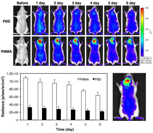
Figure 4.
Live optical imaging after PBS or PMMA implantation. P-IRDye was given via tail vein injection the following day after PBS or PMMA implantation. The mice were imaged prior and each day after the administration of the optical imaging agent for the following 6 days (n = 3). A. The upper panel shows the images from the mice with calvarial injection of PBS. The lower panel shows the images from the mice with calvarial PMMA-particle implantation. Compared to the PBS group, PMMA particle implanted animals demonstrated more intense and longer lasting NIR signals in the calvarial region where the PMMA particles were implanted; B. The NIR signal intensity was measured from a consistent region of interest (red circle) in the calvaria site for all the mice. The signal intensity differences in the two groups were statistically significant (p < 0.05).

