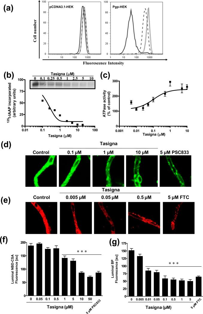
Figure 1. Tasigna inhibits Pgp-mediated efflux of calcein-AM and photoaffinity labeling of Pgp with125I-IAAP and stimulates Pgp-mediated ATP hydrolysis.
(a) pCDNA3.1-HEK and Pgp-HEK cells were incubated with 0.25 μM of calcein-AM for 10 min at 37°C in the dark, in the absence (black bold) or presence of 1 μM Tasigna (dashed line), 2 μM Tasigna (dotted line) or 2 μM Pgp specific inhibitor, XR9576 (thin line). The cells were then washed and subsequently analyzed by flow cytometer as described in the Experimental section. Representative histograms from one of three independent histograms are shown. (b) Crude membranes from High-five cells expressing Pgp were incubated with 0-10 μM Tasigna for 5 min at 21-23°C in 50 mM Tris-HCl, pH 7.5. 3-6 nM [125I]-IAAP (2200 Ci/mmole) was added and incubated for an additional 5 min under subdued light. The samples were then illuminated with a UV lamp (365 nm) for 10 min and were processed as described in the Experimental section. The incorporation of [125I]-IAAP (from autoradiogram, Y-axis) into the Pgp band was quantified by estimating the radioactivity of this band and plotted as a concentration of Tasigna (X-axis). A representative autoradiogram from one experiment is shown (inset) and similar results were obtained in two additional experiments. (c) Crude membrane protein from High-five cells expressing Pgp was incubated at 37°C with 0-10 μM Tasigna in the presence or absence of 0.3 mM sodium orthovanadate in an ATPase assay buffer for 10 min. The amount of inorganic phosphate released after incubation with 5 mM ATP for 20 min and the vanadate-sensitive ATPase activity was determined as described in the Experimental section. The average from three experiments is shown here and the error bars represent SE. (d, e) Tasigna inhibits Pgp and ABCG2 function in rat brain capillaries. Rat brain capillaries were incubated for 1 h at room temperature in the presence or absence of indicated concentrations of Tasigna or indicated inhibitors with (d) 2 μM NBD-CSA, a fluorescent cyclosporine A, which is a Pgp substrate or (e) 2 μM BODIPY® FL prazosin, a fluorescent ABCG2 substrate. Confocal images of 10 capillaries were acquired by confocal microscopy using a 40X oil immersion objective as described in the Experimental section. (f, g) Concentration-dependent decrease of lumenal NBD-CSA (f, Y-axis) or BODIPY® FL prazosin (g, Y-axis) plotted as a function of concentration of Tasigna (X-axis). Each data point represents the lumenal NBD-CSA or BODIPY® FL prazosin fluorescence mean value from 10 capillaries (pooled capillary tissue from 10 rats of a single preparation); variability is given by SEM bars. Units are arbitrary fluorescence units (au). Statistical comparison: ***, significantly lower than control capillaries, P < 0.001.

