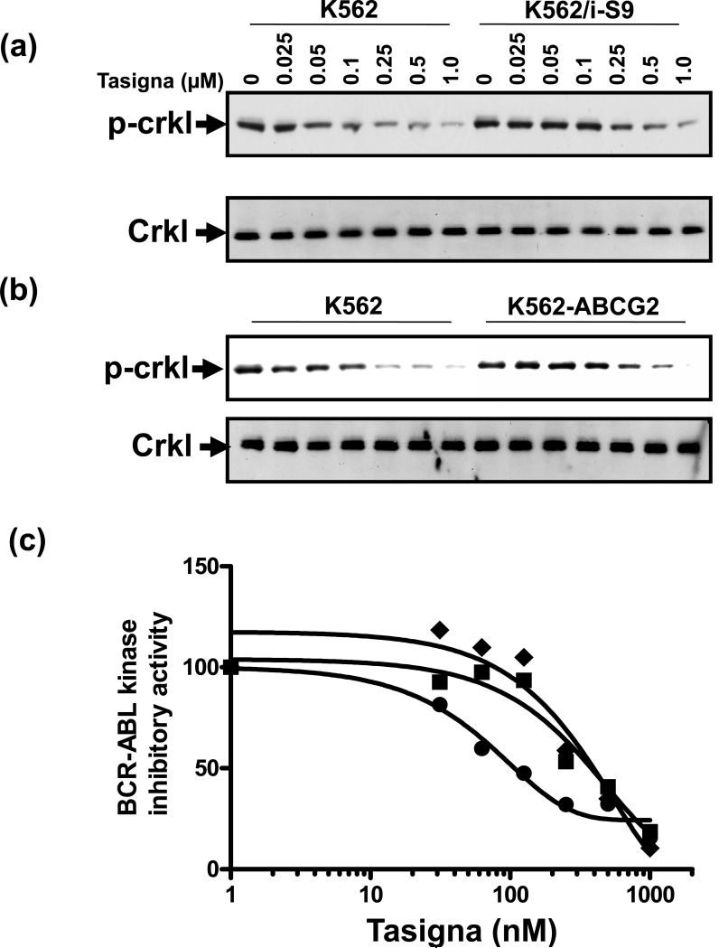
Figure 2. Effect of Tasigna on BCR-ABL kinase activity in Pgp- and ABCG2- expressing K562 cells.
K562 (lanes 1-7) in all panels, K562/i-S9 (lanes 8-14 upper panels in a) and K562-ABCG2 (lanes 8-14 upper panel in b), were incubated in the absence (lane 1, 8) or presence of indicated concentrations of Tasigna for 4 h at 37°C. The BCR-ABL kinase activity was measured by monitoring the phosphorylation of P-Crkl (upper panels in a and b) and total Crkl levels (lower panels in a and b) in cell lysates from these cells as described in the Experimental section. Shown here is a representative Western blot detecting P-Crkl and total Crkl levels from a representative experiment. Similar results were obtained in two additional experiments. (c) The P-Crkl level (measurement of BCR-ABL kinase activity) (from Western blot, Y-axis) in control K562 (●) and K562/i-S9 (■) (Pgp-expressing) or K562-ABCG2 (◆) was quantified by using the ImageQuaNT TL software and plotted as a concentration of Tasigna (X-axis).

