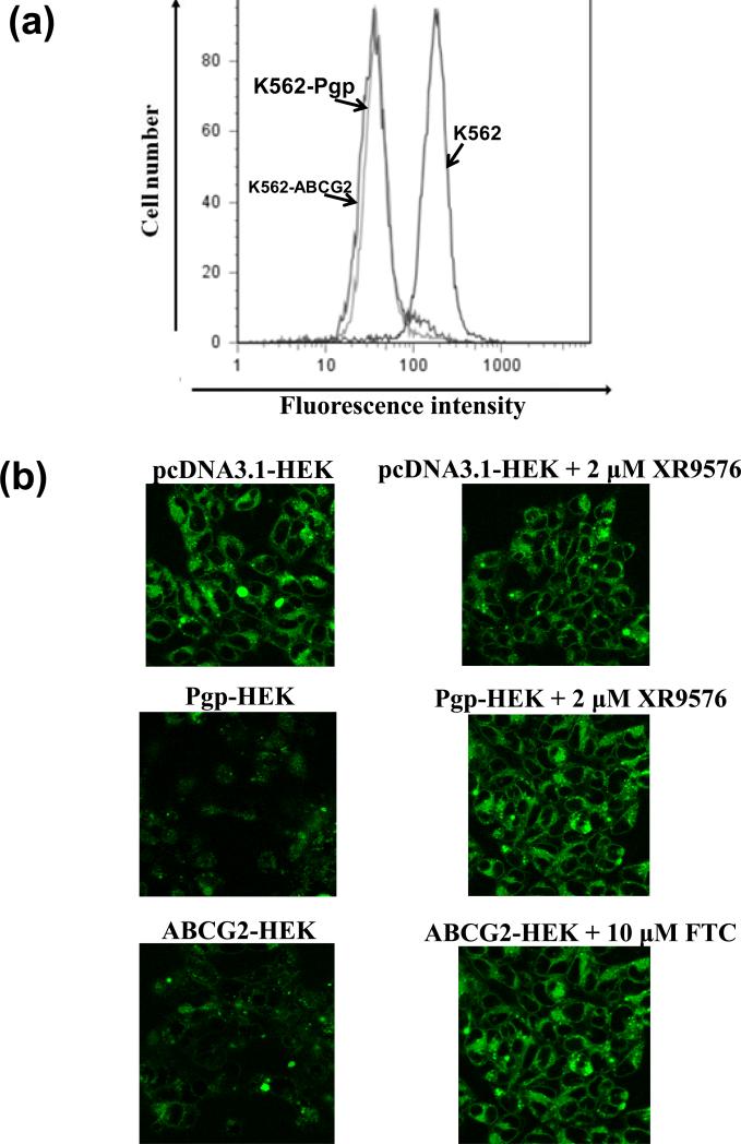
Figure 4. BODIPY® FL Tasigna is transported by both Pgp and ABCG2. (a) BODIPY® FL Tasigna is effluxed by Pgp-expressing K562 cells.
K562, K562/i-S9 (Pgp-expressing) or K562-ABCG2 (ABCG2-expressing) cells were incubated with 0.5 μM of BODIPY® FL Tasigna for 45 min at 37°C in the dark, as indicated. The cells were then washed and analyzed by flow cytometer as described in the Experimental section. Similar results were obtained in three additional experiments. (b) Detection of cellular accumulation of BODIPY®-FL Tasigna by confocal microscopy: pcDNA3.1-HEK, Pgp-HEK and ABCG2-HEK cells were incubated with 0.5 μM BODIPY® FL Tasigna in absence (left panels) or presence (right panels) of 2 μM Pgp inhibitor XR9576 or 10 μM ABCG2 inhibitor FTC at 37°C for 45 min. The cells were washed twice with cold PBS and the images were acquired as described in the Experimental section. Shown here are representative images from one experiment. Similar results were obtained in three additional experiments.

