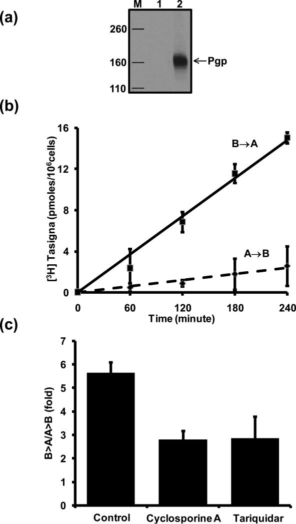
Figure 6. Transepithelial transport of [3H]-Tasigna across monolayers formed by LLC-PK1 cells expressing human Pgp.
(a) Expression of human Pgp in LLC-PK1 cells. Total cell lysates from LLC-PK1 (lane1) and LLC-PK1-MDR1 (lane 2) cells were separated by SDS-PAGE. Pgp level was detected with the anti-Pgp (C219) antibody as described in the Experimental Section. Lane M, MW markers (Kda). (b) Efflux of [3H]-Tasigna by Pgp as a function of time. [3H]-Tasigna (64 nM) was added to either the apical or basolateral side of the monolayer. An aliquot (50 μl) of the media was removed from the opposite side of the cell monolayer at indicated time points and processed as described in the Experimental section. The squares show the basolateral-to-apical (B→A) transport, the diamonds show the apical-to-basolateral transport (A→B). (c) Effect of Pgp-specific inhibitors on transepithelial transport of [3H]-Tasigna. LLC-PK1-Pgp Cell monolayers were incubated with [3H]-Tasigna (64 nM; 1 μCi/ml) in the presence or absence of cyclosporine A (10 μM) and tariquidar (1 μM) for 180 minutes. Directional drug transport ratios across cell monolayers were measured. Each point shows the mean ± SE from at least three independent experiments.

