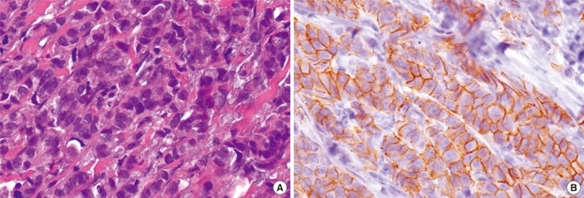
Figure 3.
(A) Microscopic findings of both breast revealed infiltration of malignant ductal cells. The primary invasive ductal carcinomas of breast shows well differentiation and no lymphovascular invasions (H&E stain, ×200). (B) The malignant cells are positive for E-cadherin (immunohistochemical staining, ×200).

