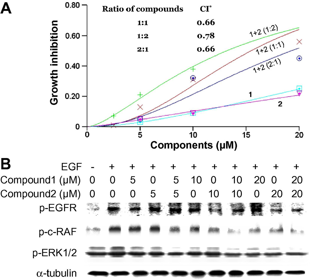
Fig. 4.
The combination of compounds 1 and 2 act synergistically to inhibit cancer cell growth. A, Growth inhibition of the 83-01-82CA cell line by individual (0, 5, 10 and 20 µM) and combined concentrations (ratio of 1:1, 1:2, and 2:1) of compounds 1 and 2. Cells were incubated for 72 h and relative cell numbers were determined by methylene blue staining. Values are normalized to vehicle DMSO controls. A combination index (CI) was determined using Calcusyn software (inset). * Combination results are synergistic when CI is <1. B, The 83-01-82CA cells were seeded and starved as in Fig. 3B, followed by incubation with compounds 1 and 2 for 1 h using the same concentration scheme (1:1) as in Fig. 4A and then incubated with 10 ng/ml EGF for 10 min. Proteins were harvested and protein levels of phosphorylated forms of EGFR (Tyr 1173) (p-EGFR), c-RAF (Ser 338) (p-c-RAF) and ERK1/2 (Thr202/Tyr204) (p-ERK1/2) were determined by Western blotting. α-Tubulin was used as a loading control.

