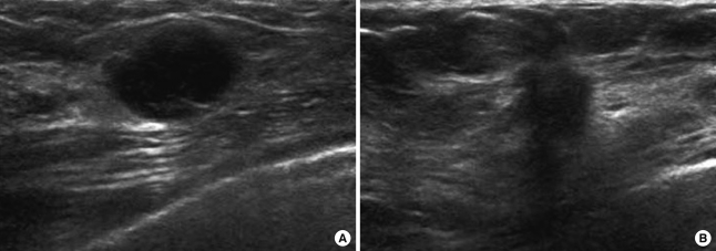
Figure 1.
Ultrasonographic findings. (A) The image of a triple receptor-negative invasive ductal carcinoma in a 30-year-old-woman. It shows an oval shaped and well circumscribed markedly hypoechoic mass without posterior shadowing. (B) The image of a non-TRN (ER+/PR+/HER2-) invasive ductal carcinoma in a 47-year-old-woman. It shows irregular shaped hypoechoic mass with spiculated margin and posterior shadowing.

