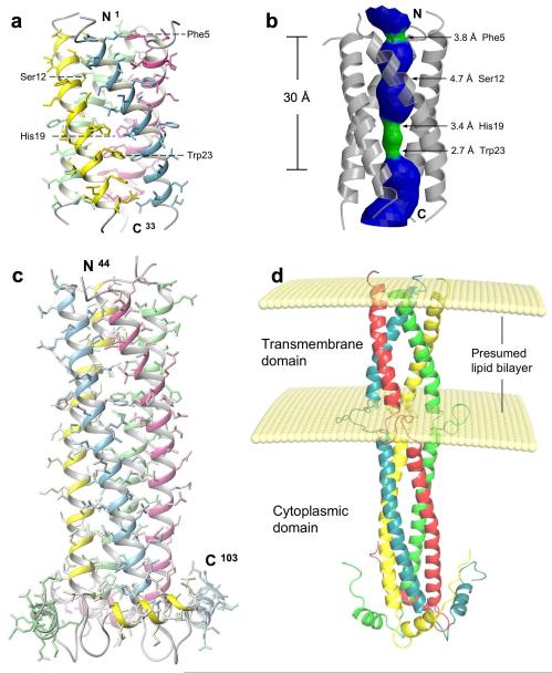
Figure 1. Solution structure of BM2 from influenza B virus.
a, Structure of the channel domain (residues 1-33) in DHPC micelles at pH 7.5. b, The pore surface of the channel domain calculated using the program HOLE 26. The channel is coloured in green for regions wider than 2.7 Å and narrower than 4.6 Å, and coloured in blue for regions wider than 4.6 Å. c, The structured cytoplasmic domain (residues 44-103) of BM2(26-109) anchored to LMPG micelles at pH 6.8. d, A model of the full-length BM2 built with the structures of the channel and cytoplasmic domains. The positioning of the two domains and their localization relative to the lipid bilayer were modeled using intra-subunit and protein-LMPG NOEs, measured for residues 29 – 43 of the BM2(26-109) construct.

