Figure 3.
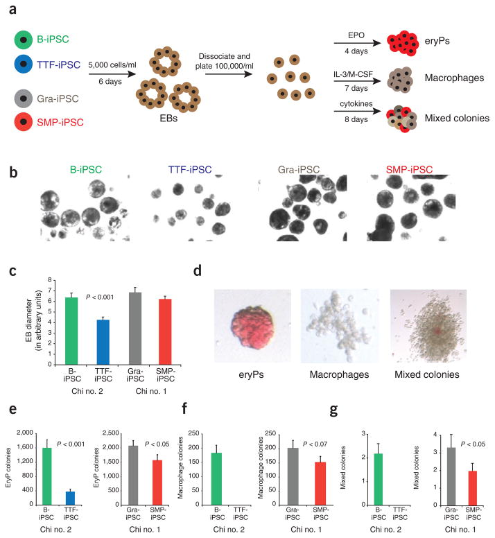
iPSCs derived from different cell types have distinctive in vitro differentiation potentials. (a) experimental outline. iPSCs were first differentiated into embryoid bodies. At day 6, embryoid bodies were dissociated and plated in conditions to favor differentiation into erythrocyte progenitors (eryP) and macrophage and mixed hematopoietic colonies. (b) Phase contrast images showing embryoid bodies derived from B-iPSCs, TTF-iPSCs, Gra-iPSCs and SMP-iPSCs at same magnification. (c) Quantification of embryoid body sizes derived from B-iPSCs, TTF-iPSCs, Gra-iPSCs and SMP-iPSCs; the diameter of the embryoid bodies was measured using arbitrary units (AU). The error bars depict the s.e.m. (n = 30) (d) Representative images of erythrocyte progenitors (eryPs), macrophage colonies and mixed hematopoietic colonies. (e–g) Quantification of in vitro differentiation potentials of the different iPSCs into EryPs (e), macrophage colonies (f) and mixed hematopoietic colonies (g). Chi no. 1, chimera no. 1; chi no. 2, chimera no. 2. The error bars depict the s.e.m. (n = 12).

