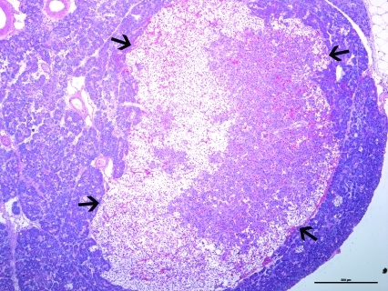
Figure 2.
Photomicrograph of the pancreatic mass from the ferret described in Figure 1. A well-delineated (arrows), expansile, moderately cellular neoplasm composed of nests and packets of neoplastic cells in a fine fibrovascular stroma was present. Hematoxylin and eosin stain; bar, 500 µm.

