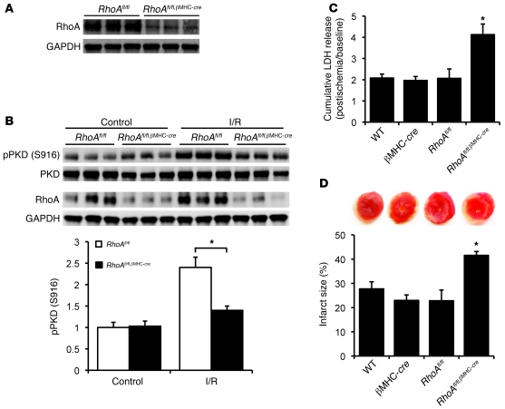
Figure 8. Cardiac-specific RhoA knockout enhances I/R injury in the heart.
(A) Representative blots showing RhoA expression in RhoAfl/fl and RhoAfl/fl,βMHC-cre hearts. (B) Representative blots and quantification showing PKD autophosphorylation in RhoAfl/fl and RhoAfl/fl,βMHC-cre hearts following I/R. *P < 0.05 (n = 3–4). (C and D) WT, βMHC-cre, RhoAfl/fl, and RhoAfl/fl,βMHC-cre hearts were subjected to global I/R. (C) LDH release to the coronary effluent following 45-minute reperfusion. *P < 0.05 versus controls (n = 4–5). (D) Representative TTC-stained ventricular sections (top); quantitative analysis of infarct size (bottom). *P < 0.05 (n = 4–7). Data are shown as mean ± SEM.

