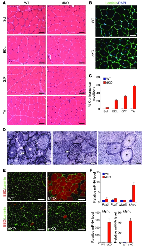
Figure 2. Centronuclear myofibers in dKO skeletal muscle.
(A) H&E staining of soleus, EDL, G/P, and TA muscles of WT and dKO mice at 12 weeks of age. Scale bars: 40 μm. (B) Immunostaining of TA muscle against laminin. Nuclei are stained with DAPI. dKO TA muscle showed central nuclei. Scale bars: 40 μm. (C) Percentage of centronuclear myofibers in 4 WT mice and 10 dKO mice at 12 weeks of age. For each mouse, more than 500 myofibers were counted for TA and G/P muscles and more than 300 myofibers were counted for soleus and EDL muscles. (D) NADH-TR staining of dKO TA muscle revealed abnormal distribution, radiating intermyofibrillary network (arrows), and ring-like fibers (asterisks). Scale bars: 20 μm. (E) EBD uptake of TA muscles of WT, dKO, and mdx mice. Immunostaining with laminin (green) is shown; EBD is detected as a red signal under fluorescence microscopy. Scale bars: 100 μm. (F) Expression of myogenic genes and of embryonic MHC (Myh3) and perinatal MHC (Myh8) in WT and dKO TA muscle, determined by real-time RT-PCR. n = 3 (WT and dKO).

