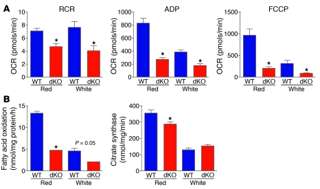
Figure 4. Mitochondrial dysfunction in dKO muscle.
(A) Mitochondria were isolated from red and white gastrocnemius muscle, and oxygen consumption rate (OCR) was measured for RCR, ADP-stimulated state 3 respiration (ADP), and FCCP-stimulated respiration (FCCP). n = 6 (WT and dKO). *P < 0.05 vs. WT. (B) Fatty acid oxidation was measured in isolated mitochondria from red and white gastrocnemius muscle. Citrate synthase enzyme activity was measured in isolated mitochondria from red and white quadriceps muscle. n = 6 (WT and dKO). *P < 0.05 vs. WT.

