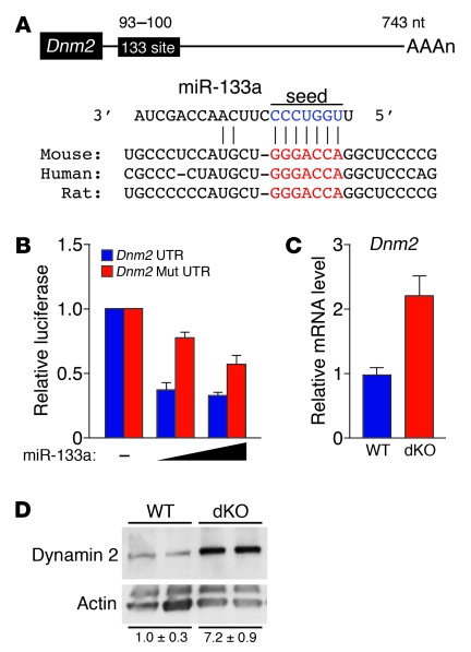
Figure 5. miR-133a regulates Dnm2 expression in skeletal muscle.
(A) Position of miR-133a target site in Dnm2 3′ UTR and sequence alignment of miR-133a and the Dnm2 3′ UTR from various species are shown. Conserved miR-133a binding sites in Dnm2 3′ UTR are shown in red. Mutations in Dnm2 3′ UTR were introduced to disrupt base-pairing with miR-133a seed sequences (blue). (B) Luciferase reporter constructs containing WT and mutant Dnm2 3′ UTR sequences were cotransfected into COS-1 cells with a plasmid expressing miR-133a. 48 hours after transfection, luciferase activity was measured and normalized to β-galactosidase activity. (C) Real-time RT-PCR showing expression of Dnm2 mRNA in WT and dKO TA muscle. n = 3 (WT and dKO). (D) Western blot showing expression of dynamin 2 protein in TA muscle of WT and dKO mice. n = 2 (WT and dKO). The blot was stripped and reprobed with an antibody against α-actin as a loading control. Quantification of dynamin 2 protein, determined by densitometry and normalized to α-actin, is also shown.

