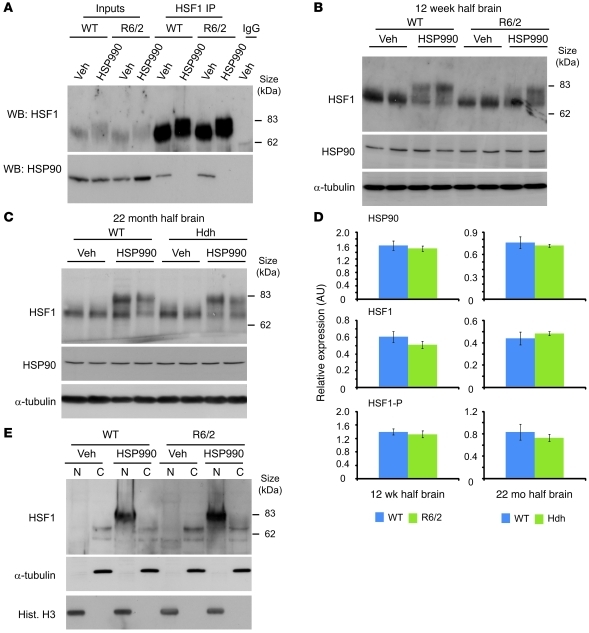
Figure 5. HSF1 dissociates from HSP90, becomes hyperphosphorylated, and translocates to the nucleus upon HSP990 treatment.
(A) Western blots of HSF1 and HSP90 after HSF1 IP from 12-week-old WT and R6/2 mouse brains 2 hours after treatment with vehicle or HSP990 (12 mg/kg). (B and C) Representative Western blots of HSF1 and HSP90 in (B) 12-week-old WT and R6/2 or (C) 22-month-old WT and HdhQ150/Q150 mouse half brains 2 hours after treatment with vehicle or HSP990 (12 mg/kg). α-Tubulin was used as a loading control. (D) Expression of HSP90 and HSF1 relative to α-tubulin, or phosphorylated HSF1 (HSF1-P) relative to unphosphorylated HSF1, in 12-week-old WT and R6/2 or 22-month-old WT and HdhQ150/Q150 mice. Densitometry values are mean ± SEM (n = 4 per treatment group). (E) Representative Western blots for HSF1 in nuclear and cytoplasmic fractions derived from 12-week-old WT and R6/2 mouse half brains 2 hours after treatment with vehicle or HSP990 (12 mg/kg). Purity of nuclear (N) and cytoplasmic (C) fractions was demonstrated by immunoblotting for α-tubulin and histone H3.

