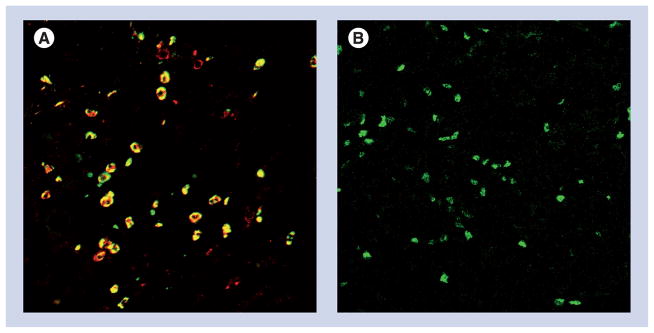
Figure 3. Immunohistology of human pancreatic adenocarcinoma.
(A) Tissue slides were stained with antibodies for CD11b (red), and CD15 (green), and shows dual positive cells (yellow), indicative of MDSC. (B) Intracellular Foxp3 staining as a marker of Treg (green). No immune infiltrate was detectable in normal human pancreas tissue (data not shown).
MDSC: Myeloid-derived suppressor cell.
Images courtesy of David C Linehan and Jonathan B Mitchem, Washington University, Department of Surgery.

