
Figure 1.
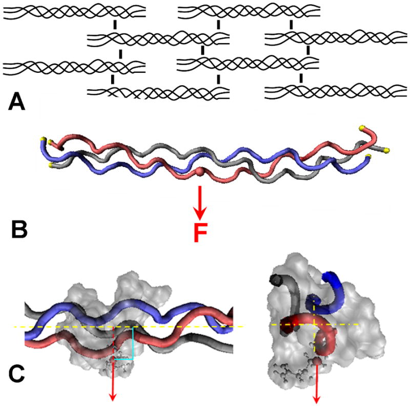
Schematic of the collagen network and location of applied force to a single chain.
(A) Simplified illustration of the collagen fibril network and the intermolecular cross-links between adjacent collagen molecules. (B) Steered Molecular Dynamics (SMD) model of the load applied to a single side chain. The C-α of each residue at the ends of the molecule were fixed in space, indicated with yellow spheres. The force, F, was applied to the C-β of an arginine residue (pseudo cross-link) through a theoretical spring (not shown). The force moved at a constant velocity in the direction indicated by the arrow. (C) Magnified view of the force applied to the C-β of the side chain, perpendicular and away from the long axis of the collagen molecule.