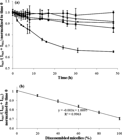
Figure 2.
(a) Stability of FRET-micelles in the presence of FBS is compared to PBS (control) and each of the major serum proteins. Time traces of the FRET ratio, I565/(I565 + I501), normalized to time 0, in solutions of (●) PBS at pH 7.4, (◼) α-, β-globulins at 15 mg/mL, (▲) γ-globulins at 15 mg/mL, (×) BSA at 45 mg/mL, and (⧫) 100% FBS (n = 3 independent experiments, mean ± standard deviation plotted). (b) Standard curve correlates FRET ratio to percentage of disassembled micelles. It is assumed that all FRET-micelles are completely disassembled after incubating with 100% FBS for 48 h (n = 3 independent experiments, mean ± standard deviation plotted).

